Cleft lip and palate are common congenital birth defects where the lip or roof of the mouth (palate) does not fully form during fetal development. These conditions occur when the tissues that normally fuse in early pregnancy fail to join properly, resulting in a gap or split. This blog draws on recent, reputable evidence to explain what cleft lip and palate are, how common they are in India, signs to watch for, causes and risk factors, how they’re diagnosed, related conditions, when to seek help, and the impact of early intervention, education, and daily life on affected individuals. The goal is to provide clear, practical, and compassionate information for families, educators, and communities. Cleft lip and palate reflect structural differences that affect feeding, speech, hearing, and appearance. The conditions vary in severity, from minor notches to complete splits, and require coordinated care across multiple specialties. Core features are present at birth and evolve with surgical and therapeutic supports throughout childhood.
Estimating cleft lip and palate prevalence in India is tricky because large, standardized studies are limited. But data suggests:

Newborns with cleft lip show a clear split in the upper lip, from a small notch to full separation into the nostril. Cleft palate appears as an opening in the roof of the mouth, sometimes isolated without lip involvement. These physical gaps are immediately noticeable at birth and affect initial feeding attempts. Parents or midwives typically identify them during the first exam. Early photos help track progress over repairs.
Babies struggle to latch due to the gap preventing airtight seal during sucking. Formula or breast milk often comes out the nose, causing choking or inadequate nutrition. Weight gain lags without special feeders; frequent small feeds become necessary. Nasal regurgitation signals palate involvement even if lip looks intact. Parental anxiety rises from these early feeding failures.
Palate dysfunction blocks Eustachian tubes, trapping fluid and inviting infections. Infants fuss, rub ears, or show delayed babbling from muffled hearing. Up to 90% experience this by age 2 without intervention. Hearing tests reveal conductive loss impacting speech sounds. Tubes prevent long-term damage if placed early.
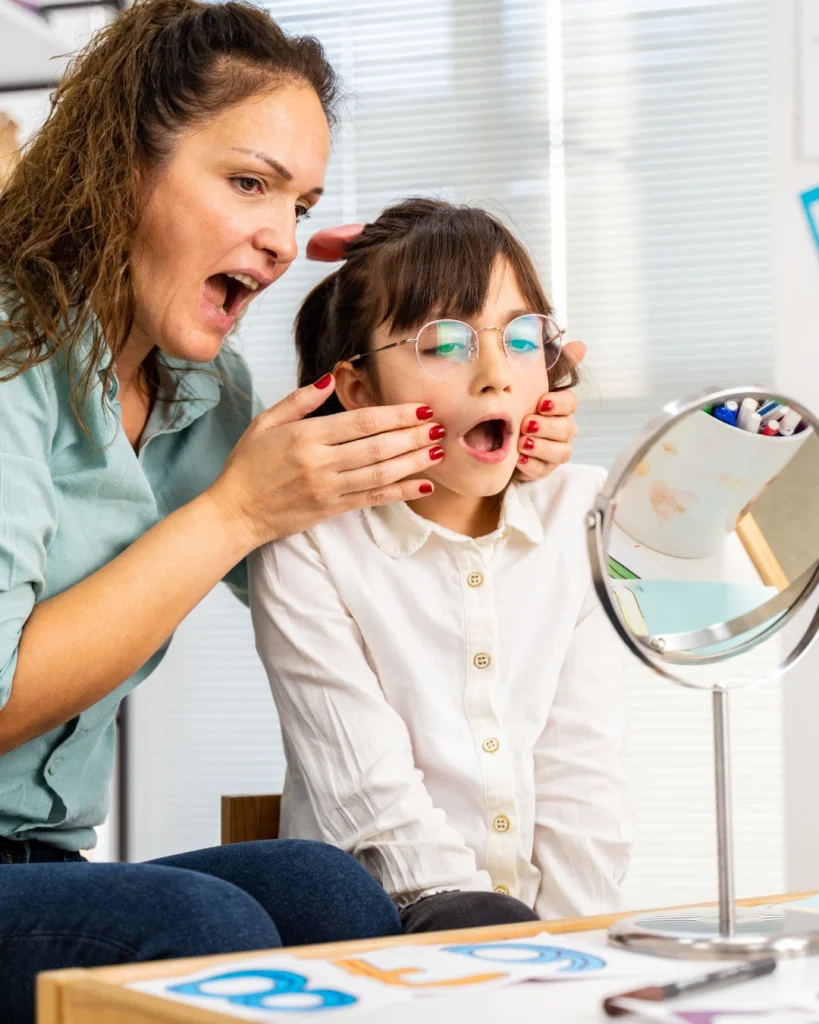
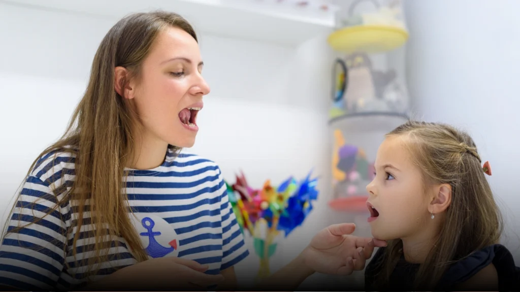
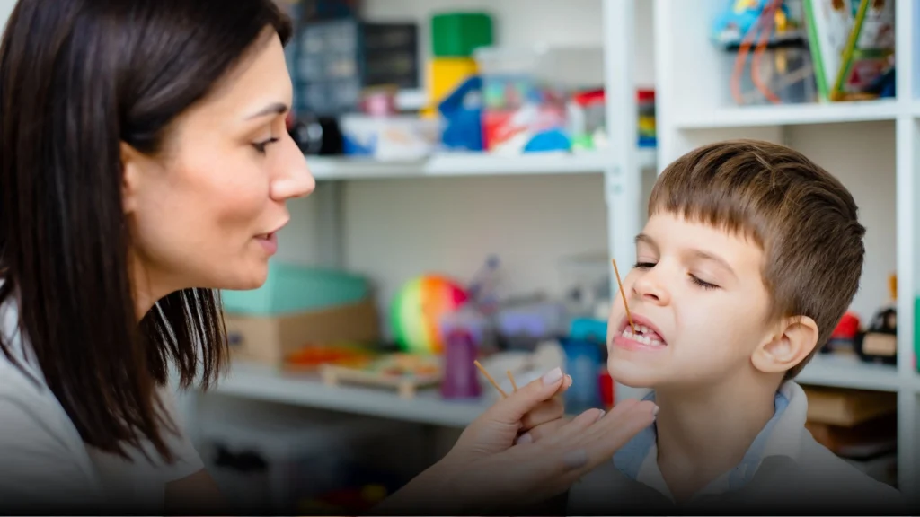
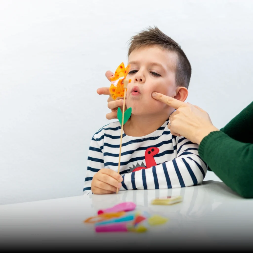
Hypernasal voice emerges as air escapes through the nose during talking. Toddlers produce muffled consonants or rely on gestures over words. Sounds like “p” or “b” distort into nasal tones without palate closure. Delays mimic language disorders until evaluated. Speech evolves dramatically post-repair with therapy.
Gaps in gums lead to missing, extra, or rotated teeth as jaws grow unevenly. Upper jaw underdevelops, causing bite issues by school age. Supernumerary teeth fill cleft spaces abnormally. Orthodontists spot this via X-rays around age 5-7. Early alignment prevents severe orthodontic needs later.
Uneven lip or nose shape persists until repairs, with one nostril wider or collapsed. Jaw growth lags on the cleft side over time. Subtle imbalances affect smiles or profiles noticeably by toddlerhood. Sequential surgeries correct these progressively. Families notice changes with each procedure milestone.
Cleft lip and palate arise from genetic factors disrupting facial fusion in weeks 4-7 of pregnancy. Over 500 genes contribute via complex interactions, not a single mutation. These affect tissue migration and signaling for lip/palate closure. Polygenic risk scores help predict familial patterns accurately. Genetics explain 70-80% of isolated cleft cases.
Siblings of affected children face 3-5% recurrence odds, rising to 10% for identical twins. Parental cleft history elevates child risk to 4-6%. Shared genes interact with environment for expression. Counseling maps inheritance patterns reliably. Most families see isolated cases without broad patterns.
Preterm delivery heightens cleft odds by interrupting critical growth phases. Babies under 37 weeks show 1.5-2x higher rates in registries. Associated low oxygen affects palate shelves. NICU monitoring flags associated defects early. Full-term pregnancies via prenatal care mitigate this.
Infants below 2.5kg carry double the cleft risk from growth restriction. Placental issues starve facial tissues of nutrients. Small size correlates with palate-only clefts especially. Postnatal weight recovery supports healing. Maternal nutrition preconception counters this factor.
Mothers over 35 or fathers over 40 show 1.2-1.5x elevated risks per studies. Accumulated mutations in eggs/sperm disrupt embryogenesis slightly. Absolute increase remains low at population levels. Fertility treatments amplify via multiples. Younger parental age offers minor protective edge.
Maternal smoking doubles cleft odds via vascular toxins halting tissue join. Alcohol, anticonvulsants, or retinoids act as teratogens too. Folic acid antagonists like methotrexate triple risks. Combined exposures compound genetically vulnerable fetuses. Avoidance preconception slashes modifiable factors.

Diagnosis typically involves a comprehensive physical evaluation by a trained professional team, including plastic surgeons, ENT specialists, orthodontists, speech-language pathologists, and audiologists. Prenatal ultrasound screens guide toward full assessment, which examines cleft extent, associated anomalies, and surgical feasibility via imaging. Diagnostic criteria follow international standards like those from the American Cleft Palate-Craniofacial Association, with India advancing universal newborn exams, multidisciplinary clinics, and protocols for early holistic management to optimize lifelong outcomes.
Conductive hearing impairment from otitis media affects 50-80% due to tube dysfunction. Chronic fluid muffles sounds, delaying language. Myringotomy tubes restore hearing in 90% cases promptly. Annual audiology prevents irreversible deficits. Behavioral signs like inattention signal needs early.
Velopharyngeal incompetence post-repair causes nasal emissions in 20%. Hypernasality persists without secondary surgery. Articulation therapy refines fricatives and stops. Multidisciplinary follow-up achieves near-normal speech by age 7. Compensatory errors embed if untreated timely.
Orofacial cleft disrupts 60% with hypodontia, peg laterals, or fistulas. Alveolar gaps demand bone grafts by age 9-11. Orthognathic surgery corrects Class III malocclusion. Prosthetics fill unrepaired defects functionally. Lifelong dental surveillance ensures alignment.
Seek evaluation if newborns show feeding struggles, nasal milk return, or visible oral gaps at birth. Refer to cleft teams by 1-2 weeks for feeding aids and surgical planning, especially if ear tugging or growth stalls emerge. Early action unlocks staged repairs, therapies, and family supports that enhance speech, hearing, and facial harmony significantly.

Specialized bottles and obturators secure suction pre-lip repair at 3 months. Consistent calories fuel immune readiness for palate surgery. Parental coaching prevents aspiration pneumonia effectively. Growth curves normalize rapidly with aids. Long-term nutrition sets surgical success foundations.
Presurgical oral motor exercises strengthen velum function early. Post-palate therapy at 12 months curbs nasal habits promptly. Alternative communication like signs bridges gaps temporarily. Auditory-verbal therapy leverages restored hearing. Verbal fluency rivals peers by kindergarten typically.
Ventilating tubes by 6 months drain effusions proactively. Prophylactic antibiotics curb recurrent media. Hearing preservation safeguards phonological development. Fewer absences improve school readiness notably. Audiograms guide precise timing interventions.
Presurgical nasoalveolar molding aligns segments before lip repair. Orthopedics minimize scar contracture effects. Sequential bone grafts enable tooth eruption. Jaw surgeries at teens perfect symmetry. Aesthetic and functional harmony emerges progressively.
Cleft lip/palate is caused by parental sins or curses.
These result from genetic and environmental factors during fetal development, not moral failings.
Kids with cleft never speak clearly
Timely surgeries plus therapy yield 90-95% normal speech; untreated cases alone cause permanent issues.
A single operation cures cleft completely
Care spans 5-7 procedures from infancy to teens, with ongoing dental/speech support for full function.
Cleft only impacts looks, not health
It causes feeding failure, chronic infections, hearing loss, and growth delays without intervention.
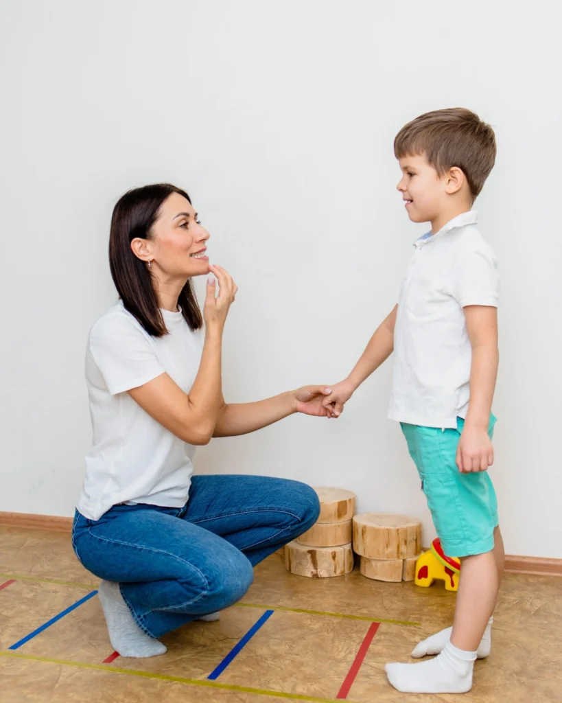
Inclusive education and accessible supports enable children with cleft lip and palate to participate meaningfully in classrooms, develop social skills, and reach academic goals. This requires trained teachers, individualized education plans, reasonable accommodations, and supportive peers.
Everyday life benefits from predictable routines, sensory-friendly environments, clear communication, and collaboration among families, schools, healthcare providers, and community services.
Societal awareness and acceptance reduce stigma and increase opportunities for employment, independent living, and community participation, reflecting a more inclusive approach to structural differences.
Seek early screening and a comprehensive evaluation if concerns arise; engage with a multidisciplinary team to craft an individualized plan.
Build routines, use visual supports and clear communication, and tailor expectations to the child’s strengths and needs.
Connect with local cleft organizations, support groups, and educational services to access resources, therapies, and advocacy opportunities.
Prioritize mental health for the whole family; caregiver stress can impact the child’s progress, so seek respite and professional support when needed.

Facial processes fail to fuse by pregnancy week 7 due to genetic variants, folate lack, or teratogens like smoking. No single trigger exists; polygenic risks combine with environment. Daily 400mcg folic acid preconception cuts incidence 20-30% reliably.
Routine 18-20 week ultrasounds detect 75% of cleft lips, less for isolated palates without profile views. 3D imaging boosts accuracy to 90%; amniocentesis checks syndromes. Preparation eases parental shock and plans delivery logistics.
Lip closure at 3 months, hard palate at 9-12 months for speech, alveolar graft ages 8-11 for teeth. Secondary rhinoplasty and jaw work follow in teens. Protocols prioritize growth windows for optimal results universally.
Yes, 70-80% need braces from age 7 to correct rotations, gaps, and bite. Bone grafts enable canine eruption properly. Coordinated teams minimize extractions and surgeries effectively.
Ear fluid causes temporary conductive loss in 80%, fixable by tubes—no IQ impact occurs. Speech lags mimic cognitive delays falsely. Interventions preserve all developmental domains fully.
Squeeze bottles like Haberman or Mead Johnson create pressure without suction needs. Upright positioning avoids reflux; thicken feeds if aspirating. Lactation consultants train parents swiftly for weight goals.
Multistage care normalizes speech in 92%, appearance in 95% per global data. Indian NGOs like Smile Train report similar via 10M+ interventions. Compliance predicts elite outcomes consistently.
Velopharyngeal issues affect 15%, resolvable surgically; rare fistulas recur under 5%. Annual multidisciplinary checks avert escalations. Most thrive without ongoing issues.
Siblings carry 4% chance, children of affected 3-6%; twins near 40% concordant. Genetic tests refine odds precisely. Folate and avoidance lower modifiable risks.